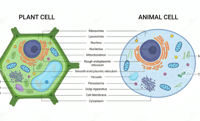
The animal cell diagram serves as an essential visual resource for grasping the fundamental building blocks of animal life. It illustrates the intricate internal organization of eukaryotic cells found in humans, animals, and other multicellular organisms. By highlighting specialized structures known as organelles, the diagram reveals how these tiny units perform vital tasks such as energy generation and waste removal. For those pursuing general knowledge in biology, an transforms abstract concepts into clear, relatable visuals that foster deeper appreciation of cellular biology. This guide explores its key elements to make learning both informative and engaging.
The Basic Layout of an Animal Cell Diagram
A typical animal cell diagram presents the cell in an irregular, rounded shape without the rigid cell wall seen in plants. The plasma membrane forms the outer boundary, acting as a dynamic barrier that selectively allows nutrients and signals to pass while expelling waste. Inside lies the cytoplasm, a fluid matrix that suspends organelles and facilitates chemical reactions essential for survival. This flexible structure, as depicted in the enables cells to change shape for movement or engulfing particles through processes like endocytosis. Such visuals underscore why animal cells adapt efficiently to diverse physiological demands across tissues and organs.
The Nucleus: Control Center in the Animal Cell Diagram
Prominently featured in every animal cell diagram is the nucleus, often the largest organelle and easily identifiable by its double membrane. It houses the cell’s genetic material in the form of chromatin, which condenses into chromosomes during division. The nucleolus within the nucleus assembles ribosomes, crucial for protein production. This central role makes the nucleus the command hub directing cellular activities like growth and reproduction. Studying its placement in the helps explain how genetic instructions coordinate everything from metabolism to response to environmental changes.
Mitochondria: Powerhouses Highlighted in the Animal Cell Diagram
Mitochondria appear as bean-shaped structures with inner folds called cristae in the animal cell diagram, emphasizing their role in energy production. These organelles convert nutrients into adenosine triphosphate through cellular respiration, supplying the power needed for all cellular functions. Their own DNA and ribosomes suggest an ancient bacterial origin, adding evolutionary depth to their depiction. The abundance of mitochondria in high-energy cells, such as muscle or nerve cells, is often noted in diagrams to illustrate efficiency. This feature clarifies why disruptions in mitochondrial function link to various health conditions.
Endoplasmic Reticulum and Ribosomes in the Animal Cell Diagram
The animal cell diagram clearly shows the endoplasmic reticulum as a network of interconnected membranes extending from the nucleus. Rough endoplasmic reticulum, studded with ribosomes, specializes in synthesizing and folding proteins destined for export or membrane insertion. Smooth endoplasmic reticulum handles lipid production and detoxification without ribosomes. Ribosomes themselves, tiny dots either free in the cytoplasm or attached, translate genetic code into proteins. Together, these components demonstrate the cell’s sophisticated manufacturing system, making the invaluable for understanding protein pathways.
Golgi Apparatus and Lysosomes Depicted in the Animal Cell Diagram
The Golgi apparatus stands out in the animal cell diagram as stacked, flattened sacs near the nucleus, responsible for modifying, sorting, and packaging proteins and lipids into vesicles. It acts like a cellular post office, directing materials to their destinations inside or outside the cell. Lysosomes, shown as small vesicles containing digestive enzymes, break down worn-out organelles and engulfed particles. Their acidic interior ensures safe waste processing. These elements in the illustrate the cell’s efficient recycling and distribution mechanisms, preventing buildup of harmful substances.
Centrioles and Cytoskeleton Supporting the Animal Cell Diagram
Near the nucleus, the often includes a pair of centrioles within the centrosome, essential for organizing microtubules during cell division. The cytoskeleton, a network of protein fibers, provides structural support, enables movement, and transports organelles. This framework allows the cell to maintain shape while permitting dynamic changes. In the these features highlight how animal cells achieve coordinated division and intracellular transport without a rigid wall. Such details reveal the remarkable engineering that supports tissue formation and repair in living organisms.
FAQs
What is the primary purpose of an animal cell diagram?
An visually maps the locations and relationships of organelles, making it easier to comprehend how cells maintain life processes. It aids students and enthusiasts in identifying structures at a glance and connecting them to specific functions like energy production or protein synthesis. This clarity supports better retention of biological concepts and prepares learners for advanced topics in physiology and genetics.
How does an animal cell diagram differ from a plant cell diagram?
An lacks a cell wall, chloroplasts, and a large central vacuole, reflecting the absence of photosynthesis and rigid support in animal cells. Instead, it emphasizes centrioles for division and lysosomes for digestion. These distinctions in the highlight evolutionary adaptations, helping viewers quickly recognize why animal cells prioritize flexibility and heterotrophic nutrition over plant-like rigidity.
Why are mitochondria so important in the animal cell diagram?
Mitochondria receive special attention in the because they generate most of the cell’s usable energy through respiration. Their detailed structure, including folded membranes, underscores efficiency in high-demand tissues. Disruptions here can lead to fatigue or disease, so the diagram reinforces their critical role in overall cellular health and organism survival.
What role do centrioles play as shown in the animal cell diagram?
Centrioles appear in the as cylindrical structures that organize the spindle fibers during mitosis, ensuring accurate chromosome separation. This function is vital for growth, repair, and reproduction. Their presence distinguishes animal cells and explains efficient division processes, providing key insights into developmental biology and potential abnormalities.
Conclusion
In summary, the animal cell diagram offers a clear window into the coordinated world of organelles that sustain animal life. From the plasma membrane’s selective control to the nucleus’s genetic oversight, mitochondria’s energy output, and the supportive roles of the endoplasmic reticulum, Golgi apparatus, lysosomes, and centrioles, each component works in harmony. This visual tool not only enhances general knowledge but also sparks curiosity about cellular mechanisms underlying health and disease. By mastering the, readers gain lasting appreciation for the microscopic foundations of all animal existence.